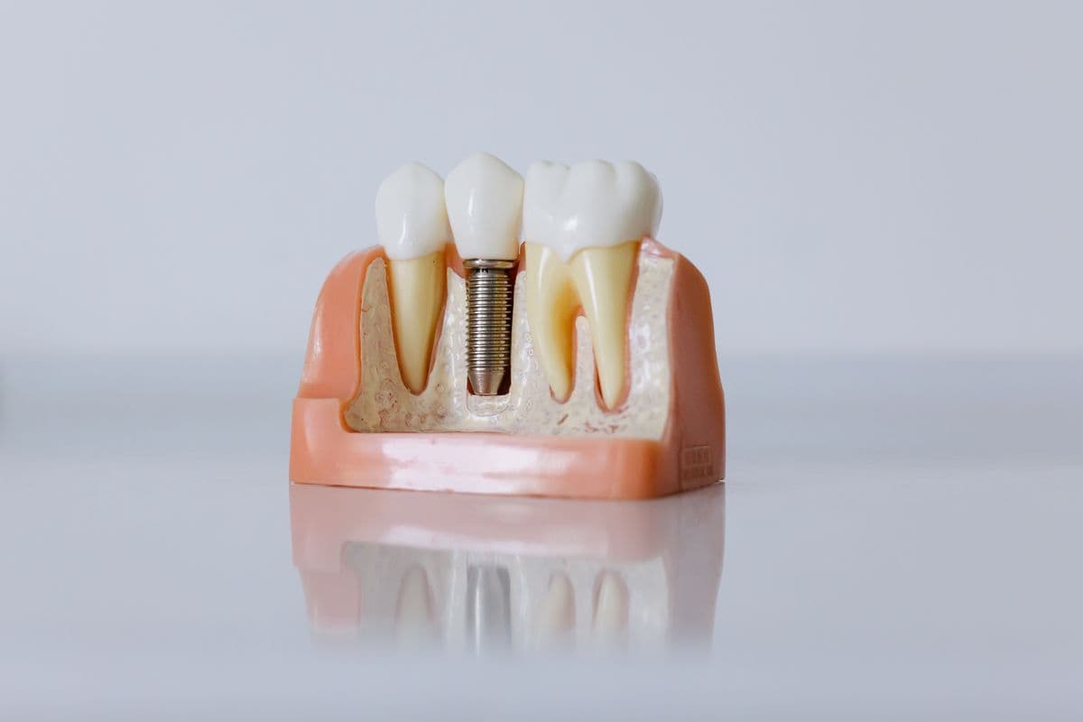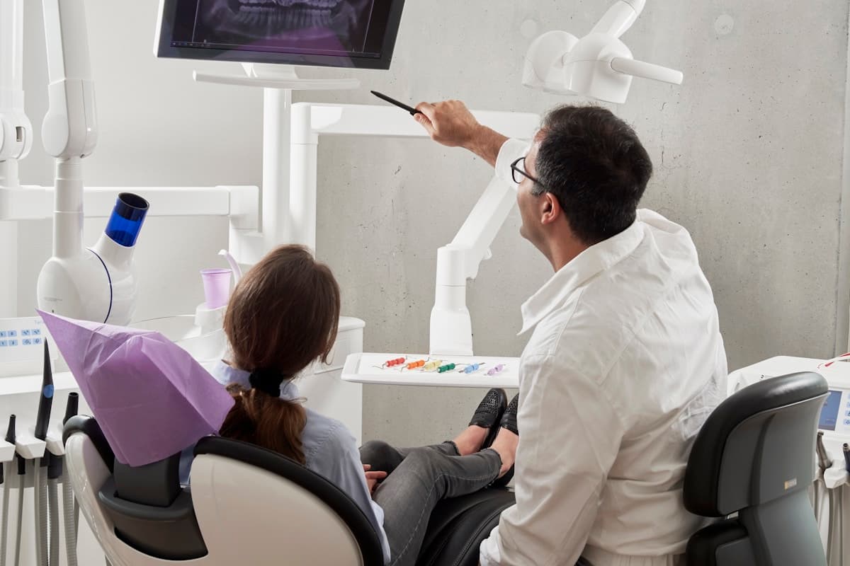
CBCT imaging transformed implant planning from two-dimensional guesswork into three-dimensional precision, but the value of a CBCT scan depends entirely on how clinicians and planners process that data into an actionable treatment plan. This article traces the complete pathway from scan acquisition through implant plan delivery, covering the technical steps our team executes when processing cases submitted via our digital workflow.
Why a Structured Workflow Matters
- Three-dimensional visualization of bone anatomy, nerve pathways, and sinus boundaries
- Accurate implant sizing and positioning based on volumetric bone assessment rather than periapical estimates
- Seamless transfer from planning data to surgical guide fabrication without manual remeasuring
- Documented planning records that support informed consent and medicolegal documentation
Where It Fits in Treatment Planning
This workflow applies whenever a CBCT-based surgical plan needs to translate cleanly into a guide or prosthetic outcome, from single-tooth cases to full-arch reconstructions.
- Posterior mandible cases requiring inferior alveolar nerve identification and safety zone planning
- Maxillary implant sites near the sinus floor where grafting or lift planning is needed
- Multi-implant cases where parallelism and distribution must be verified in three dimensions
- Immediate placement scenarios where extraction socket morphology affects implant trajectory
The Digital Workflow, Step by Step
Each step in this workflow exists to catch a specific failure point—scan distortion, registration error, or guide misfit—before it reaches the surgical stage.
- Acquire CBCT with appropriate field of view and resolution for the planned implant sites
- Segment anatomy: mandibular canal, maxillary sinus, cortical plates, and relevant pathology
- Merge intraoral scan or model scan with CBCT using radiographic stent or matching landmarks
- Position implants with prosthetic overlay and verify safety margins to critical structures
- Design and export surgical guide for printing or milling, with drilling protocol documentation

Best Practices
- Use a limited field of view CBCT centered on the implant site for optimal resolution
- Apply radiographic stents with fiduciary markers when merging CBCT with intraoral scans
- Maintain a minimum 2 mm safety margin from the inferior alveolar nerve unless documented otherwise
- Review merged data for alignment errors before committing to guide fabrication
Common Pitfalls
- Using panoramic or periapical radiographs alone for implant depth planning in complex anatomy
- Failing to verify CBCT-to-scan merge accuracy, resulting in guide misalignment at surgery
- Ignoring artifact from existing metal restorations that obscure bone assessment in CBCT
- Selecting implant length without accounting for apical bone concavity visible only in CBCT
“A digital workflow only earns its accuracy claims if every handoff between scan, plan, and guide is verified—skipping a check anywhere in the chain reintroduces the error digital planning was meant to remove.”
Conclusion
Strong outcomes in cbct to implant plan: the complete digital workflow depend on clear clinical goals, accurate records, and a planning partner who understands manufacturing requirements. Explore our specialist service, review the case submission workflow, or contact our team to discuss your next case.